Perinatal pathology is a study of the fetus, the placenta, and the neonate. Perinatal pathology department in Mediscan is the first, the oldest and the only lab dedicated exclusively for the study of fetuses, placentas and neonates in India and has been doing high quality fetal autopsies for the past 25 years. It is dedicated towards diagnosis of fetal congenital anomalies, infections, metabolic disorders and finding the cause of fetal death, growth restriction, and absence of fetal wellbeing.
Services provided by the department include fetal and neonatal autopsy, and placental pathology.
We do complete fetal / neonatal autopsy which includes
After completion of the autopsy, the x rays and organ histology are reported, all the findings are summarised, correlated with the history and antenatal and postnatal investigations, and the conclusion with recommendations are given. This includes extensive review of literature and consultations with clinical geneticists whenever necessary.
Once a week review meetings are held along with the sonologists, geneticists and the fetal medicine doctors to correlate our findings with the ultrasound findings which is a gold standard for feedback and for honing sonological skills. This has contributed towards making Mediscan a world class centre for fetal medicine and training. Reviews with clinical geneticists adds valuable information regarding advice to be given to the parents and is incorporated in the report.
This kind of detailed workup helps in collating all the possible information about the fetus which is essential for counselling the parents regarding subsequent pregnancies, and aids the doctors involved in planning the right type of care for the mother, the unborn, and the neonate, and provides continuous feedback to sonologists which is invaluable for skill improvement and cannot be provided in such a comprehensive manner by any other modality.
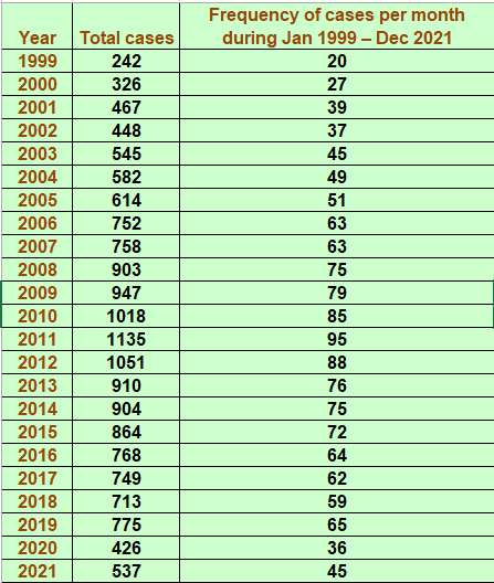
Has been in existence for more than 25 years doing exclusive perinatal pathology work and is the largest of its kind in India. Till date more than 15000 autopsies have been conducted successfully and reports given to the parents.
The department received NABL accreditation in AUGUST 2021
We are not part of a hospital and so all the fetuses that we get are sent to us from outside by obstetricians attached to various hospitals in Chennai and from other cities in Tamil Nadu and in other states. The fetus and placenta have to be immersed in 10% formalin and sent in watertight container to the department.

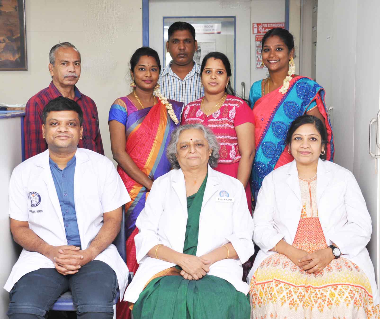
Dr Lata is the senior most consultant and was trained in pathology at PGIMER Chandigarh. She is a dedicated and passionate perinatal pathologist who has been doing exclusive perinatal work for the past 25 years in Mediscan. She started the department and has built it to a world class setup. Her areas of special interest are congenital cardiac anomalies and skeletal dysplasias.

Dr.Poornima is a consultant and was trained in pathology at PG institute of basic medical sciences, Taramani,Chennai.She has been associated with mediscan for the past 16 years and was trained by Dr.Lata at Mediscan.Her areas of special interest are fetal radiology & congenital CNS anomalies.

Dr Harish is a trained pathologist and has been with mediscan since 2017 doing exclusive perinatal pathology work. He is an active participant in doing autopsies and in all the acedemic activities of the department and the institution. He especially enjoys teaching and is specially interested in cardiac dissection.
We accept fetus from GA of 11 weeks till term and neonates
All fetuses should be ideally sent along with the placenta and the whole length of cord attached to the placenta
Specimen should be accompanied by a detailed history which includes the marital, and obstetric history, history of consanguinity of parents and grandparents, history of any congenital anomaly or pregnancy loss in the family including siblings of the parents, maternal medical history before and during the present and past pregnancies, and reports of relevant investigations done before and during the pregnancy.
Details should include whether the pregnancy was terminated or if the miscarriage was spontaneous, the status of the fetus before delivery, whether the fetus was stillborn or liveborn, all the ultrasound reports done during the pregnancy, the GA of the fetus at the time of delivery, and the date of the LMP. If the conception was assisted, that should be mentioned and also the type of intervention done e.g., IVF, ICSI.
Points to note while sending the fetus
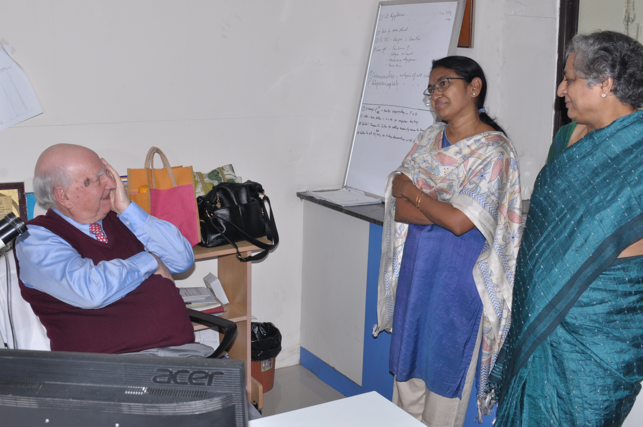
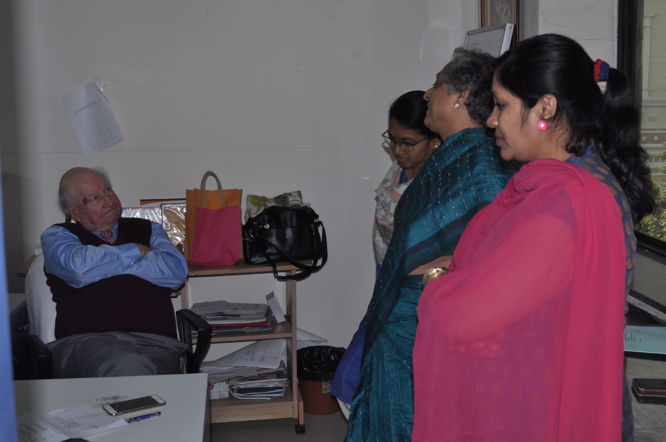
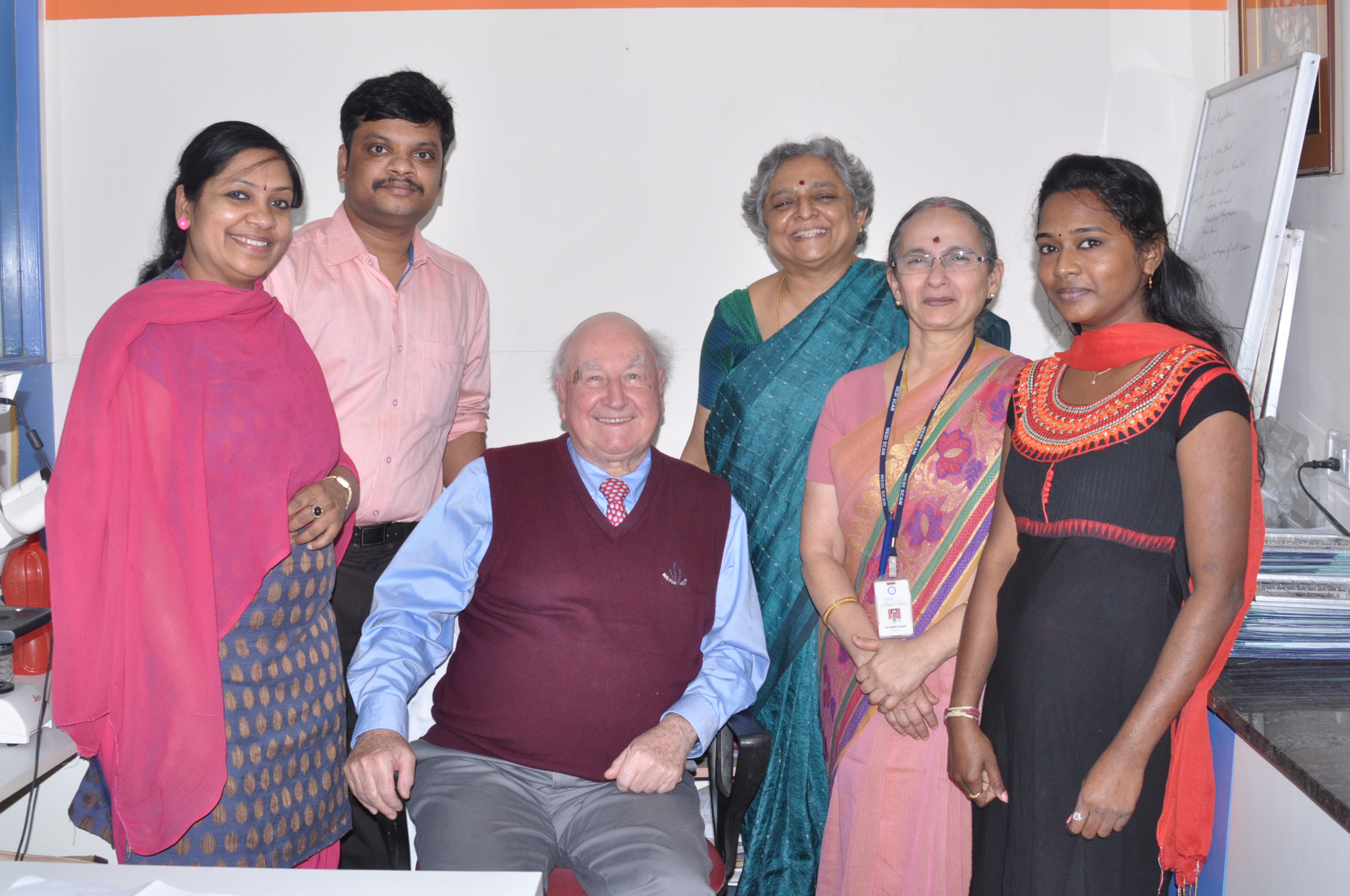
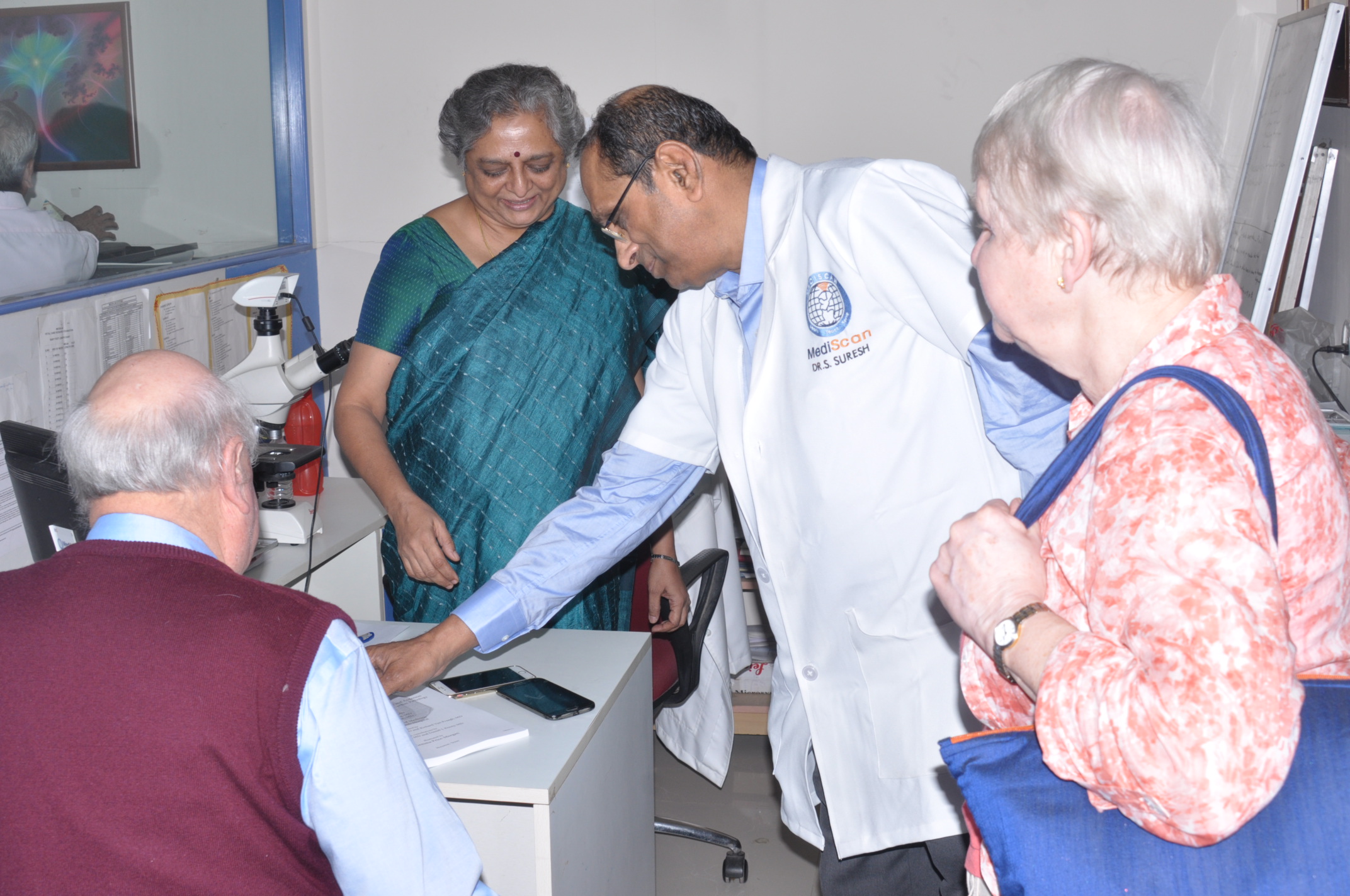
International Faculty visit :
January 2018
Prof.Robert H Anderson
Cardiac Morphologist & International faculty
United Kingdom
In perinatal pathology the fetus and the placenta are examined in detail. The study includes – external examination, examination of all the organs both by naked eye and under the microscope, and the x ray of the baby for study of the fetal skeleton.
There are three experienced pathologists in the department who study the baby in detail and give a report.
The examination is done after termination of the pregnancy for a severe congenital abnormality identified during pregnancy (on ultrasound) , or when we need to know the possible cause of demise when the baby in inside the uterus (intrauterine demise)
Although the fetus has been examined on ultrasound during pregnancy, since the ultrasound scans cannot identify all the anomalies and the complete extent of the anomalies. If the fetus has died during the pregnancy, the cause of death can be determined in many cases only on examination of the fetus and the placenta.
Only if all the abnormalities in the fetus are identified, a final diagnosis can be arrived at. This ,will help plan further investigations when necessary. With the results one can know whether the condition is likely to recur in subsequent pregnancies or not. This knowledge is essential for the parents and the treating doctor(s) both for reassurance and planning for future pregnancies.
Ideally it should be done soon after delivery. As this may not be possible, one should bring the baby as early as possible. If there is a delay they or the baby is delivered at a time when the department is closed, the baby is preserved in a preservative fluid as described below in point no 8
When the baby is in the womb, its organs are not yet fully developed and cannot perform all the functions necessary for the baby to survive. The baby gets its oxygen, nutrition, and some essentials needed for the wellbeing from the mother through the placenta. If there is some abnormality in the placenta this will be affected and the baby will not thrive. Many of the abnormalities in the placenta can be seen only on examining the tissue under the microscope. Similarly, the umbilical cord and the membranes within which the fetus is enclosed may also show abnormalities. These can be identified only when we do a proper pathological study of the entire placenta. Not examining the placenta will result in missing crucial information.
The doctor or the nurse (in the place of delivery) should wash the baby and immerse it completely in formalin solution (10%). The container should be large, and have a watertight lid. The lid should be further sealed with tape and the container should have a label with the necessary information. The baby should be brought with the medical history and all the investigations done during the pregnancy. All the scan and blood reports must be there. A requisition letter from the doctor stating the reason for requesting the autopsy is a must.
Preferably the father of the child or an authorized person with due request form and consent form and ID proof of the father / mother.
While giving the baby for a pathological examination, an official of the department will ask some essential questions about the pregnancy, the mother’s health, and family history. The answers to these are crucial for a giving the report. Also, the examination can be done ONLY with a signed consent from one of the parents. The consent form will be given to the father for signing in the department.
The cost at present is Rs. 6500.00 but it may be revised in the future and so you can call the department to find out.
In about 6 months. The department will call and inform you when the report is ready.
It can be handed over to the parent or sent by courier whichever is desired by the parents.
For collecting the report in person one of the parents should come (we cannot hand over the report to others for privacy and legal reasons) and bring the yellow card given by the department at the time of handing over the fetus. This card has the department number which is required for handing over the report. This number should be quoted for any enquiry regarding the report or the baby.
The Pathology Department primarily helps in analysis of fetuses with abnormalities which have been terminated. They help to answer questions related to fetal abnormality or death.
a. The fetus / placenta is registered with an I.D. number. If the mother has already been registered in Mediscan, it is advisable for her to bring the ID card issued to her previously. This will facilitate better analysis and reporting of abnormal findings at autopsy.
b. A detailed history of the mother is obtained - past medical history / pregnancy related problems/ drugs consumed during pregnancy etc.
c. The family history of the couple is elicited.
d. All scans and other medical reports of the mother are recorded.
e. Consent forms authorizing the Department to proceed with analysis of the fetus, have to be signed.
a. X - rays of the fetus are taken
b. The fetus is examined in detail externally - head to foot, all possible and relevant measurements are recorded.
c. The internal organs are then examined in detail.
d. All relevant and necessary tissues are examined under the microscope for more details.
a. A report consists of a detailed explanation of what was normal / abnormal in the fetus.
b. All the abnormalities are listed
c. These findings are put together to consider the various causes and their effects.
d. Thus a complete diagnosis is arrived at wherever possible and it is reported.
e. Where a confirmed diagnosis has not been made, the closes possible conditions are enlisted.
a. The report of the pathology examination is used by the geneticist/ dysmorphologist / obstetrician to counsel the couple regarding the recurrence risks of the problem in subsequent pregnancy
b. If an antenatal ultrasound has shown abnormalities, the pathology examination gives additional information in more than 30% of the patients.
c. The need for further testing for the couple can be assessed
Yes, from the Genetic counseling Department, provided the referring doctor has asked for it. Hence, a written request for the same is essential. A prior appointment from the Genetic counseling Department has to be obtained.
The cleaned fetus / placenta have to be preserved in 10 % formalin for testing. (10 % formalin is obtained by mixing 1 part formalin with 10 parts of water).
It should be completely immersed in this liquid.
The container should be of adequate size to preserve the fetus well.
If the fetus is preserved well, the chances of finding answers are better.
Once the autopsy is completed, the Fetal medicine experts, Geneticists and Pathologists have a weekly review meeting in house, wherein each report is reviewed and the next plan of action is charted. This information is provided to the couple if they seek our consultation . In case they opt out of the discussion, then the report is despatched as per the couple’s wish